B-Pestaña-1-Artículo-May-18-2019_1.jpg

May 18, 2019 English Blog

The goal of this clinical trial was to assess the feasibility and safety of transplanting autologous bone marrow mononuclear cells into patients suffering severe embolic stroke. Major inclusion criteria included patients with cerebral embolism, age 20-75 years, National Institute of Health Stroke Scale (NIHSS) score displaying improvement of ≤ 5 points during the first 7 days after stroke, and NIHSS score of ≥ 10 on day 7 after stroke. Bone marrow aspiration (25 or 50 mL; N = 6 patients in each case) was performed 7-10 days poststroke, and bone marrow mononuclear cells were administrated intravenously. Mean total transplanted cell numbers were 2.5 × 10(8) and 3.4 × 10(8) cells in the lower and higher dose groups, respectively. No apparent adverse effects of administering bone marrow cells were observed. Compared with the lower dose, patients receiving the higher dose of bone marrow cells displayed a trend toward improved neurologic outcomes. Compared with 1 month after treatment, patients receiving cell therapy displayed a trend toward improved cerebral blood flow and metabolic rate of oxygen consumption 6 months after treatment. In comparison with historical controls, patients receiving cell therapy had significantly better neurologic outcomes. Our results indicated that intravenous transplantation of autologous bone marrow mononuclear cells is safe and feasible. Positive results and trends favoring neurologic recovery and improvement in cerebral blood flow and metabolism by cell therapy underscore the relevance of larger scale randomized controlled trials using this approach.

Click here to read the full article


B-Pestaña-1-Artículo-May-2-2019_1-1.jpg

May 2, 2019 English Blog

City doctors have used stem cells to save the life of a premature baby boy, who was suffering from a chronic lung disease. When all efforts failed to revive Rudransh Dubey’s lungs, who was born at 26 weeks (normal gestation period is 39 weeks), the doctors decided to try the stem cell therapy, which is in an experimental stage. They injected the baby with 40 million stem cells and gradually the lungs began to repair, and he could be weaned off the ventilator, continuous positive airway pressure (CPAP) machine, and oxygen support step by step.

Rudransh was born on June 27 last year at a Malad hospital and he weighed merely 600 grams. “Premature babies like him have very tiny air sacks. Breathing for them thus becomes very difficult,” said neonatologist Dr. Nandkishore Kabra from Surya Hospital in Santacruz where Rudransh was shifted a month after his birth.

According to Dr. Kabra, the baby was put on all the possible lines of treatment like steroids, vitamins, and surfactant to develop the lungs and wean him off the ventilator support. However, his condition remained critical and he continued to require high-pressure ventilator support.

Rudransh had developed severe broncho pulmonary dysplasia. While in most cases, babies in such conditions lose their battle for life, in Rudransh’s case, the doctors and his parents decided to try the stem cell therapy as a last attempt. Third party stem cells derived from umbilical cord were sourced by Rudransh’s parents. The doctors injected about 40 million cells directly into the baby’s lungs through the intratracheal tube.

“About 10 days later, the baby’s ventilator support was going down. The lungs had begun to develop better,” Dr. Kabra said. Gradually, the baby was shifted on CPAP machine and then later on simple oxygen support with which he was discharged from the hospital on March 11. He weighed 4.6 kg at the time of discharge. “For the last few weeks, the baby has completely been off any breathing support,” he said.

Doctors said stem cells were used as a life-saving measure. “This therapy is not a standard of care. We have to exercise caution,” paediatrician Dr. Bhupendra Avasthi from Surya Hospital said.

He said the procedure was carried out free of cost. “The consent of the parents and the hospital’s ethics committee was taken, and the Declaration of Helsinki that provides guidance on medical research was followed to carry out the procedure,” Dr. Avasthi said.

Dr. Kabra said intra-pulmonary stem cell therapy in such a chronic lung disease has never been used before. “Researchers in Korea have used mesenchymal stem cells in babies as a prophylactic while researchers in Australia have used amnion stem cells intravenously in babies to see their safety. However, we have gone a step further and used stem cells in a baby as the last available therapy,” he said.

Rudransh’s parents refused to divulge the source from where they obtained the stem cells.

I kept on pushing the doctor to find an alternative. I have enormous faith in science and I know science does new things everyday,” said Pramod.


B-Pestaña-1-Artículo-April-26-2019_1-1.jpg

April 26, 2019 English Blog

Doctors at Loma Linda University Children’s Hospital recently conducted the institution’s first stem cell transplant in a sickle cell disease patient, effectively curing her of the inherited blood disease. The successful procedure procedure offers hope and accessible treatment to those suffering from the disease in the Inland Empire and surrounding regions.

Children’s Hospital doctors had worked for nearly a year to build a program focused on helping hematology patients, specifically hemophilia and sickle cell disease.

Akshat Jain, MD, pediatric physician specializing in hematologic disorders at Children’s Hospital, said he is pleased with the outcome of the transplant and what it means for future patients suffering from sickle cell disease.

“We created a successful program so children and their families suffering from this disease don’t need to look elsewhere for treatment — it’s available to them right here in the Inland Empire,” Jain said.

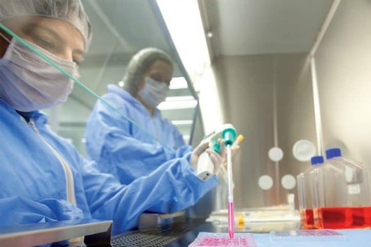
The procedure was also Children’s Hospital’s first haploidentical transplant, meaning the stem cells donated — by the patient’s father — were only half a genomic match to the patient’s own stem cells. The transplant team infused the father’s cells directly into the patient after conditioning chemotherapy to replace the unhealthy blood-forming cells..

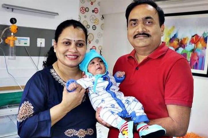
The patient, 11-year-old Valeria Vargas-Olmedo, had lived with sickle cell disease since birth. Her family began seeking treatment last year after she became incapacitated, unable to continue daily activities such as attend school, get in a car or even walk. Doctors said she had debilitating chronic pain, bone loss and bone necrosis.

“She is now disease free and can go back out into the world to do what an 11-year-old should be doing,” Jain said.

Sickle cell disease causes a shortage of red blood cells and thus an oxygen deficiency in one’s body. This can cause chronic pain and other serious complications, such as infection, acute chest syndrome and stroke. Without oxygen, any organ has a high likelihood of dying off.

Jain said the disease is generally found in populations like those in the Inland Empire, such as Hispanic and African American populations.

Jain said he and his team treat approximately 250 to 300 sickle cell patients in Children’s Hospital’s comprehensive sickle cell program — more patients than in some of the largest programs on the west coast.

Clara Olmedo, Valerie’s mother, said, “Firstly, we want to thank God. We also want to thank Dr. Jain and his entire transplant team. Finally, thanks to Valerie’s father — he did everything he could in order to save her life and give her health through being a donor. My daughter is much more animated now — she’s begun walking, she’s eating and gaining weight, she’s happy. Little by little she is living a normal life like before.”

The Vargas-Olmedo family wants to encourage others families who are struggling with sickle cell disease. “For the parents who see the news of this transplant and deal with this sickness, I hope they are encouraged and know that Children’s Hospital is a great hospital,” Clara Olmedo said. “There are many good doctors, professionals and excellent nurses. I encourage them to ask more questions about this procedure and our experience. They’ve helped us tremendously, and we have our trust in them.”


B-Pestaña-1-Artículo-February-14-2019_1.jpg

February 14, 2019 English Blog

Background:  The underlying pathophysiology in intellectual disability  (ID) involves  abnormalities in dendritic branching and connectivity of the neuronal network. This limits the ability of the brain to process information. Conceptually, cellular therapy through its neurorestorative and neuroregenerative properties can counteract these pathogenetic  mechanisms and improve neuronal connectivity.  This improved networking should exhibit as clinical efficacy in patients with ID.

Methods:  To assess the safety and efficacy of cellular therapy in patients with ID, we conducted an open-label proof-of- concept study from October 2011 to December 2015. Patients were divided into two groups: intervention group (n = 29) and rehabilitation group (n = 29). The intervention group underwent cellular transplantation consisting of intrathecal administration of autologous bone marrow mononuclear cells and standard neurorehabilitation. The rehabilitation group underwent only standard neurorehabilitation.

The results of the symptomatic outcomes were compared between the two groups. In the intervention group analysis, the outcome measures used were the intelligence quotient (IQ) and the Wee Functional Independence Measure (Wee-FIM). To compare the pre-intervention and post-intervention results, statistical analysis was done using Wilcoxon’s matched-pairs test for Wee-FIM scores and McNemar’s test for symptomatic improvements and IQ. The effect of age and severity of the disorder were assessed for their impact on the outcome of intervention. Positron emission tomography- computed tomography (PET-CT) brain scan was used as a monitoring tool to study effects of the intervention. Adverse events were monitored for the safety of cellular therapy.

Results: On symptomatic analysis, greater improvements were seen in the intervention group as compared to the rehabilitation group. In the intervention group, the symptomatic improvements, IQ and Wee-FIM were statistically significant.  A significantly  better outcome of the intervention was found in the paediatric age group (<18 years) and patients with milder severity of ID. Repeat PET-CT scan in three patients of the intervention group showed improved metabolism in the frontal, parietal cortex, thalamus, mesial temporal structures and cerebellum. No major adverse events were witnessed.

Conclusions: Cellular transplantation with neurorehabilitation is safe and effective for the treatment of underlying brain deficits in ID.

Click here to read the full article


B-Pestaña-1-Artículo-February-11-2019_1.jpg

February 11, 2019 English Blog

Cellular therapy is an emerging therapeutic modality with a great potential for the treatment of autism. Recent findings show that the major underlying pathogenetic mechanisms of autism are hypoperfusion and immune alterations in the brain. So conceptually, cellular therapy which facilitates counteractive processes of improving perfusion by angiogenesis and balancing inflammation by immune regulation would exhibit beneficial clinical effects in patients with autism. This is an open label proof of concept study of autologous bone marrow mononuclear cells (BMMNCs) intrathecal transplantation in 32 patients with autism followed by multidisciplinary therapies. All patients were followed up for 26 months (mean 12.7). Outcome measures used were ISAA, CGI, and FIM/Wee-FIM scales. Positron Emission Tomography-Computed Tomography (PET-CT) scan recorded objective changes. Out of 32 patients, a total of 29 (91%) patients improved on total ISAA scores and 20 patients (62%) showed decreased severity on CGI-I. The difference between pre- and postscores was statistically significant () on Wilcoxon matched-pairs signed rank test. On CGI-II 96% of patients showed global improvement. The efficacy was measured on CGI-III efficacy index. Few adverse events including seizures in three patients were controlled with medications. The encouraging results of this leading clinical study provide future directions for application of cellular therapy in autism.

Click here to read the full article


B-Pestaña-1-Artículo-February-4-2019_1.jpg

February 4, 2019 English Blog

AIM:

Management of osteoarthritis (OA) is basically symptomatic. Recently, stem cells (SC) have been used in the search for an optimum treatment. We decided to conduct a controlled clinical trial to determine if a single intra-articular injection of in vivo stimulated bone marrow SC could lead to an improvement in pain management and quality of life in patients with knee OA.

METHOD:

This was a prospective, open-label, phase I/II clinical trial to assess the safety and efficacy of a single intra-articular injection of autologous stimulated bone marrow stem cells (BM-SC) in patients with knee OA. Individuals of both genders older than 30 years with confirmed diagnosis of OA who signed informed consent were included in two groups: SC group received in vivo BM stimulation with subcutaneous administration of granulocyte colony stimulating factor (G-CSF). SC were obtained by BM aspiration and administered in a single intra-articular injection. The control group received exclusively oral acetaminophen. Visual analogue scale and Western Ontario and McMaster Universities Osteoarthritis Index scores were performed at 1 week, 1 month and 6 months in both groups. This trial was registered in ClinialTrials.gov NCT01485198.

RESULTS:

A total of 61 patients were included. Socio-demographic characteristics, OA grades and initial scores were similar in both groups. The BM-SC group showed significant improvement in knee pain and quality of life during the 6-month follow-up.

CONCLUSION:

The study demonstrates feasibility and supports efficacy of a completely ambulatory procedure in treatment of knee OA.

Click here to read the full article


B-Pestaña-1-Artículo-January-28-2019_1.jpg

January 28, 2019 English Blog

BACKGROUND:

Quality of life (QOL) is an important factor in evaluating the effectiveness of treatment in children with cerebral palsy (CP). The aim of this study was to evaluate the effects of autologous bone marrow mononuclear cells (BM MNCs) on the QOL of children with CP.

METHODS:

From December 2015 to December 2016, 30 children with CP aged from 2 to 15 years received two intrathecal infusions of BM MNCs, one at baseline and the other 3 months later, at Vinmec International Hospital. The motor function and muscle tone of the patients were evaluated using the Gross Motor Function Measure (GMFM)-88 and Modified Ashworth Score, respectively. Their QOL was assessed at baseline and 6 months after the first BM MNC transplant using the Vietnamese version of the Cerebral Palsy Quality of Life Questionnaire for children (CP QOL-Child)-the parental proxy report, which comprises seven domains. Nineteen mothers (mean age: 32.9±4.9 years) and 11 fathers (mean age: 36.1±6.8 years) were invited to complete the CP QOL-Child assessment before and after the transplantations, Paired t-tests and multivariate regression analyses were used to evaluate the changes in QOL and GMFM scores and to identify the key factors correlated with the QOL score.

RESULTS:

Significant changes were observed in the children’s gross motor function and muscle spasticity, as evidenced by the GMFM-88 total score, scores for each of its domains, the GMFM-66 percentile and the muscle tone (P < 0.001). Six months after the transplantations, the QOL scores of children with CP were markedly increased (P < 0.001) for all the domains, except for the domain of access to services. In the multivariate regression analysis, significant associations were found between higher age of children and higher QOL except for feeling about functioning and pain and impact of disability domains. Gross Motor Function Classification System (GMFCS) level was negatively correlated with the score of pain and impact of disability domain, while the GMFM-88 scores were positively correlated with the QOL in terms of feelings about functioning and family health domain (P < 0.05).

CONCLUSION:

The QOL of the children with CP was noticeably improved 6 months after BM MNC transplantation and was accompanied by improvements in gross motor function and muscle tone.

TRIAL REGISTRATION:

ClinicalTrials.gov Identifier: NCT02574923 . Registered on October 14, 2015.

Click here to read the full article


B-Pestaña-1-Artículo-January-21-2019_1.jpg

January 21, 2019 English Blog

Cerebral Palsy (CP) is a disabling condition that affects a child’s life and his/her family irreversibly. It is usually a non-progressive condition but improvement over time is rarely seen. The condition can be due to prenatal hypoxia, metabolic, genetic, infectious, traumatic or other causes. It is therefore a heterogeneous group that results in functional motor disability associated with different degrees of cognitive abnormalities. There are no treatments that can cure or even improve CP and the best available approach aims at functional, social and nutritional supportive care and counseling. In this paper, we report 17 sequential patients with CP treated with intrathecal administration of Bone Marrow Mononuclear Cells (BMMC). All patients had an uneventful post-injection course with 73% of the evaluable patients treated having a good response using the Gross Motor Function Classification System (GMFCS). The average improvement was 1.3 levels on the GMFCS with cognitive improvements as well.

Click here to read the full article


B-Pestaña-1-Artículo-January-14-2019_1.jpg

January 14, 2019 English Blog

Cerebral palsy is a nonprogressive heterogeneous group of neurological disorders with a growing rate of prevalence. Recently, cellular therapy is emerging as a potential novel treatment strategy for cerebral palsy. The various mechanisms by which cellular therapy works include neuroprotection, immunomodulation, neurorestoration, and neurogenesis. We conducted an open label, nonrandomized study on 40 cases of cerebral palsy with an aim of evaluating the benefit of cellular therapy in combination with rehabilitation. These cases were administered autologous bone marrow mononuclear cells intrathecally. The follow-up was carried out at 1 week, 3 months, and 6 months after the intervention. Adverse events of the treatment were also monitored in this duration. Overall, at six months, 95% of patients showed improvements. The study population was further divided into diplegic, quadriplegic, and miscellaneous group of cerebral palsy. On statistical analysis, a significant association was established between the symptomatic improvements and cell therapy in diplegic and quadriplegic cerebral palsy. PET-CT scan done in 6 patients showed metabolic improvements in areas of the brain correlating to clinical improvements. The results of this study demonstrate that cellular therapy may accelerate the development, reduce disability, and improve the quality of life of patients with cerebral palsy.

Click here to read the full article


B-Pestaña-1-Artículo-January-7-2019_1-1200x900.jpg

January 7, 2019 English Blog

Cell therapy is being widely explored in the management of stroke and has demonstrated great potential. It has been shown to assist in the remodeling of the central nervous system by inducing neurorestorative effect through the process of angiogenesis, neurogenesis, and reduction of glial scar formation. In this study, the effect of intrathecal administration of autologous bone marrow mononuclear cells (BMMNCs) is analyzed on the recovery process of patients with chronic stroke. 24 patients diagnosed with chronic stroke were administered cell therapy, followed by multidisciplinary neurorehabilitation. They were assessed on functional independence measure (FIM) objectively, along with assessment of standing and walking balance, ambulation, and hand functions. Out of 24 patients, 12 improved in ambulation, 10 in hand functions, 6 in standing balance, and 9 in walking balance. Further factor analysis was done. Patients of the younger groups showed higher percentage of improvement in all the areas. Patients who underwent cell therapy within 2 years after the stroke showed better changes. Ischemic type of stroke had better recovery than the hemorrhagic stroke. This study demonstrates the potential of autologous BMMNCs intrathecal transplantation in improving the prognosis of functional recovery in chronic stage of stroke. Further clinical trials are recommended. This trial is registered with NCT02065778

Click here to read the full article


Bioss Stem Cells Mexico Logo

Bioss Stem Cells 2022 – All rights reserved